Introduction
A continuous layer of food-grade encapsulating material encloses solid particles, liquid droplets, or gaseous compounds in the widely used technique known as microencapsulation. The resulting capsules are usually between 1 and 1000 μm in size. This technology has been widely used in a variety of industries, such as food processing, pharmaceuticals, and agriculture (1,2). Because of their wide range of health-promoting benefits, microencapsulation has become especially relevant in the food industry for the delivery of probiotics. Probiotics have been shown to protect against rotavirus infection, relieve atopic eczema (3), rhinitis, and asthma, and have antimicrobial, anti-carcinogenic, and anti-diarrheal qualities (4). Probiotics have also been linked to several physiological benefits, including lowering serum cholesterol, modifying immune responses, and suppressing Helicobacter pylori infections (5). These characteristics highlight how crucial it is to create efficient encapsulation techniques to guarantee probiotic viability and functionality in food systems (6,7). Probiotic encapsulation is being sought to improve the resistance of probiotics to harsh conditions that may be encountered during food processing, storage, and passage through the gastrointestinal tract. Oxidation, shear force, drying, and exposure to temperature, moisture, and oxygen can significantly decrease viability, while gastric acidity and bile salts can further limit survival (8). Lactobacillus and Bifidobacterium species are currently the most well-studied probiotics because of their proven health benefits. To be effective, the International Dairy Federation requires a minimum viable number of ≥ 10^7 CFU/g at the time of consumption, emphasizing the importance of probiotic encapsulation. These factors emphasize how crucial it is to use efficient encapsulation techniques to preserve probiotic stability and functionality during processing, storage, and digestion (9).
Selection of appropriate wall material is also of great importance as it determines the physical and chemical properties of the resultant microcapsules. Several substances, including carbohydrates, proteins, and waxes, have been used as wall materials. This makes it an ideal agent of the carbohydrate-based encapsulating agents, sodium alginate is the most used. Alginates are naturally occurring anionic polysaccharides of alginic acid, which are found in large amounts in the cell walls of seaweeds and as capsular polysaccharides in some bacteria. Their biocompatibility, non-toxicity, easy gelation conditions, and low immunogenicity have made them widely used in the pharmaceutical, food, and biomedical industries (10). Sodium alginate, the salt of alginic acid, is especially useful for encapsulation because of its ability to create hydrogels in the presence of divalent ions such as calcium, barium, or strontium ions. This property makes sodium alginate an ideal encapsulating agent for probiotics. (11).
The application of prebiotics to improve the functioning of probiotics has been gaining momentum in recent years. Aloe vera (Aloe barbadensis), a natural source of prebiotics, has been identified as a promising candidate owing to its multifaceted nutritional and medicinal properties. The plant is rich in vital vitamins, minerals, amino acids, and phytochemicals like urea nitrogen, cinnamic acid, salicylic acid, phenols, lupeol, and sulphur, which possess antimicrobial properties. Specific components such as acemannan and fructans from Aloe vera have demonstrated ability to enhance Bifidobacterium growth and SCFA formation. It is because of this complex phytochemical composition that Aloe vera has been found to possess a wide range of biological properties like anti-diabetic, antimicrobial, gastroprotective, laxative, antioxidant, anti-inflammatory, anti-tumour, anti-allergic, hypoglycaemic, immunomodulatory, and wound-healing properties (12, 13).
The objective of the study was thus, to use a combination of sodium alginate and Aloe vera for encapsulation of Lactobacillus acidophilus using emulsion technique.
Materials and Methods
Materials
Procurement of Culture. Freeze- dried pure culture of Lactobacillus acidophilus 291 used in the study was purchased from National Collection of Dairy Cultures, ICAR- National Dairy Research Institute, Karnal, India.
Procurement of Reagents. Sodium alginate, calcium chloride and Tween 80 (emulsifier) required for the study were of reagent grade. Aloe vera juice and vegetable oil were purchased from local shops.
Methods
Activation of freeze- dried culture. The bacterial culture of Lactobacillus acidophilus NCDC 291 was activated by subculturing in MRS broth at 37°C for 24-48 hours in an incubator (14). A culture of 0.65 absorbance (109 Colony Forming Unit (CFU)/ml) at 600 nm was used for the purpose (15).
Preparation of cell suspension. The bacterial culture of Lactobacillus acidophilus obtained after subsequent subculturing was harvested by centrifugation at 3000 rpm at 4°C for 15 minutes. The pellet was washed and resuspended in 0.85% (w/v) saline solution for the encapsulation process (16).
Preparation for Microencapsulation of the activated culture. All the glasswares and reagents used in the procedure were sterilized.
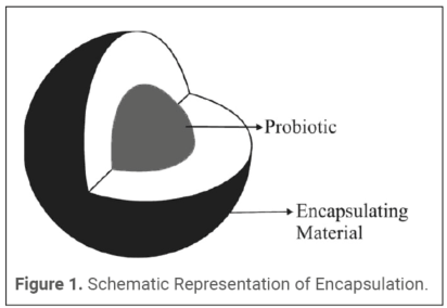
Initially, capsules were prepared using only sodium alginate as the wall material for standardization, at various concentrations of 0.5%, 1%, 1.5% and 2% (% weight/ volume) with cell suspension in volume ratio of 1:1, 1:2, 1:3, 1:4 and 1:5 (Table 1). For emulsion technique, the final cell suspension–alginate mixture mixed thoroughly using a magnetic stirrer was added to vegetable oil containing 0.2% Tween 80 emulsifier in the ratio of 1:5 and mixed thoroughly using a magnetic stirrer. Upon emulsion formation, calcium chloride solution (0.05M) was added dropwise to the turbid emulsion until the beads separated leaving a clear supernatant. The resultant calcium alginate beads were allowed to harden for 30 minutes and then filtered using sterilized Whatman No. 1 filter paper and washed with 0.85% saline solution (16). Figure 1 depicts the schematic representation of the structure of the microencapsulated particle.
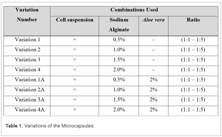
The same procedure was followed by adding 2% Aloe vera to the sodium alginate- cell suspension mixture and the encapsulation efficiency in each case was calculated.
Analysis of the Probiotic survival rate. Probiotic survival rate was determined by releasing of the entrapped probiotic from the matrix. The releasing activity was done by dissolving the beads using phosphate buffer (0.1M, pH 7.4) in a ratio of 1:9 (w/v). The phosphate ions chelate the calcium ions thereby weakening the alginate beads for effective release of the cells. The homogenised sample was serially diluted till 10-9 and cultured with MRS agar which was then incubated at 37 °C for 48 hours in anaerobic condition. Triplicates were prepared and an average was calculated. All the variations were performed ten times for obtaining appropriate results. The Probiotic survival rate was, thereafter, calculated according to the formula:
Survival rate = (N/N0) × 100
Where N is the number of viable entrapped cells released from the beads and N0 is the number of free cells added to the biopolymer mix during the encapsulation process (17).
Statistical Analysis. All experiments were performed in triplicate and data were expressed as mean ± standard deviation (SD). Appropriate statistical software was used for statistical analyses. Two-way analysis of variance (ANOVA) was used to study the effect of formulation variation and the ratio of cell suspension to wall material on the encapsulation yield. If significant differences were found, the Tukey’s honestly significant difference (HSD) post-hoc test was used to perform pairwise comparisons of the treatment means. Statistical significance was defined at p < 0.05. The coefficient of variation (CV) was also calculated to evaluate the reproducibility and consistency of the encapsulation process.
Results
Estimation of the Probiotic survival rate
Table 2 and Table 3 show the probiotic survival rate (in percentage) of all the variations performed. Table 2 displays an overall increase in probiotic survival rate with a decrease in ratio increase in concentration of sodium alginate from 0.5% to 2%. These findings are in accordance with the study conducted by Sheu and Marshall (16). The results also indicate a decrease in the encapsulation efficiency with increase in ratio from 1:1 to 1:5 in Variation 1 and 2, whereas an increase in percentage efficiency from 1:1 to 1:5 in Variations 3 and 4.
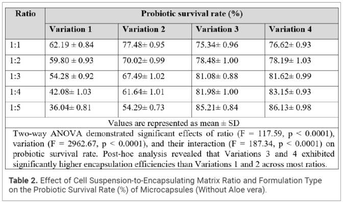
Table 3 specifies the result of probiotic survival rate after the addition of 2% Aloe vera to the cell suspension-alginate mixture. The concentration of Aloe vera was maintained at due to formation 2% as mentioned in a study conducted by Syahirah et al. in 2013 where encapsulation efficiency was found to be the highest with addition of 2% Aloe vera using FCCD-RSM approach (18). In the present study, all the variations done using sodium alginate alone were repeated in presence of Aloe vera. Results showed a decrease in the encapsulation efficiency with change in ratio from 1:1 to 1:5 in 0.5% and 1% alginate concentration whereas, an increase in the percentage of probiotic survival rate was seen from 1:1 to 1:5 ratio in variations containing 1.5% and 2% sodium alginate. In high concentration of gelling agents, formation of microcapsules was not observed, but gel was formed that was hard in structure and did not disrupt to release the entrapped cells during analysis of the probiotic survival rate. Lower percentage of probiotic survival rate was observed in Variation 4A.
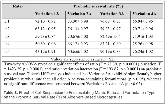
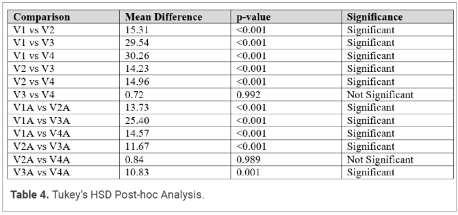
Two-way ANOVA revealed that both the encapsulation ratio and formulation significantly influenced probiotic survival rate (p < 0.001). A significant interaction effect between ratio and formulation was also observed (p < 0.001). Tukey’s HSD analysis (Table 4) showed that Variation 3A exhibited significantly higher encapsulation efficiency than all other Aloe vera-containing formulations, whereas Variations 3 and 4 of the alginate-only system were statistically comparable. The highest probiotic survival rate (90.19 ± 0.83%) was obtained with Variation 3A at a ratio of 1:5, indicating that incorporation of Aloe vera enhanced microencapsulation efficiency.
The highest probiotic survival rate of 90.19 ± 0.83% was obtained with 1.5% sodium alginate and 2% Aloe vera when the cell suspension- alginate ratio of 1:5 was used. To check for further increase or decrease in yield, 1:6 ratio combination was also carried out, and results showed a decline in the yield maybe due to decrease in viscosity of the mixture.
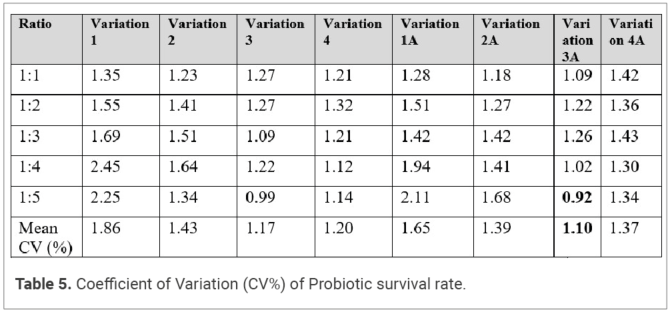
Table 5 shows the results of the Coefficient of Variation (CV) to observe the consistency of the models. The table clearly indicates that both the models are consistent. The encapsulation process exhibited low variability and excellent precision across all treatments. The addition of Aloe vera not only improved probiotic survival rate but also maintained very low variability, confirming its stabilizing role in enhancing matrix formation and encapsulation efficiency. Of all the variations, Variation 3A exhibited lowest variability (Mean CV= 1.10%) and best reproducibility among all tested formulations, particularly at ratio 1:5 (0.92%). The dataset overall demonstrates good reproducibility of the encapsulation process.
Discussion
The decrease in encapsulation efficiency with an increase in ratio from 1:1 to 1:5 in Variations 1 and 2, and the increase in encapsulation efficiency in Variations 3 and 4, may be related to differences in the physicochemical properties of the cell suspension–alginate mixtures. In Variations 3 and 4, higher ratios led to higher encapsulation efficiency under the present processing conditions, most probably due to the formation of a more stable gel network and more effective polymer-core interactions.
It has been shown in previous studies that viscosity and gel-forming properties of sodium alginate are important factors information and stability of microcapsules. The sodium alginate used in the present study was composed of guluronate and mannuronate residues, the latter being in greater proportion. Literature suggests that mannuronate-rich alginate tend to have higher solution viscosity while guluronate-rich segments are attributed to stronger gel formation by ionic cross-linking (19). Proper combinations of the components can result in beads with good elasticity and structural stability (20,21). However, as viscosity was not measured in the present study, the role of viscosity in the observed encapsulation behaviour is speculative.
Further, the effect of incorporation of Aloe vera along with sodium alginate was studied on the survival rate of probiotic. Variations containing sodium alginate and Aloe vera showed a decrease in encapsulation efficiency with increasing ratio from 1:1 to 1:5 at 0.5% and 1.0% alginate concentrations. An increase in encapsulation efficiency was observed at 1.5% and 2.0% alginate concentrations. Interestingly, some formulations with Aloe vera showed higher probiotic survival rates than those with alginate alone. These differences could be explained by different composition and structural properties of the encapsulating matrix due to different concentration of gelling agents. The lower apparent probiotic cell survival in Variation 4A may potentially be correlated with a firmer gel structure in the matrix which inhibited total entrapment cell release in encapsulation efficiency evaluation. This should be clarified with rheology and structural assessment.
Approximately 3-4% increased probiotic survivability was observed following the incorporation of Aloe vera versus respective alginate-only controls. This could be attributed to the presence of polysaccharides such as acetylated glucomannan, galactan, galactogalacturan, glucogalactomannan, galacto-glucoarabinomannan, and glucuronic acid-containing polymers that have been identified in Aloe vera gel, which may help improve encapsulation matrix structure, cell entrapment, and overall retention of encapsulated cells within the alginate beads (22).
While earlier studies have identified that Aloe vera gel can affect viscosity and gel properties, this was not assessed herein and studies including rheological measurements would be required to further explore this factor.
Conclusion
The present study demonstrated the potential of sodium alginate, both alone and in combination with Aloe vera, as effective wall materials for probiotic microencapsulation using the emulsion technique. Encapsulation yield was significantly influenced by the ratio of cell suspension to encapsulating material, the formulation composition, and the interaction between these factors. Two-way ANOVA revealed significant effects of ratio, variation, and ratio × variation interaction on encapsulation yield for both sodium alginate-only formulations and sodium alginate–Aloe vera formulations (p < 0.0001). These findings indicate that optimization of both wall material composition and formulation ratio is critical for achieving maximum encapsulation efficiency.
Among the sodium alginate-only formulations, Variations 3 and 4 exhibited significantly higher encapsulation yields than Variations 1 and 2, with no significant difference observed between Variations 3 and 4. In the sodium alginate–Aloe vera formulations, Variation 3A produced the highest encapsulation yield and was significantly superior to all other Aloe vera-containing formulations. The highest encapsulation yield obtained in the study was 90.19 ± 0.83% for Variation 3A at the 1:5 ratio, suggesting that the incorporation of Aloe vera can enhance encapsulation performance when used in appropriate proportions.
Aloe vera is recognized for its rich polysaccharide content and prebiotic potential and is widely utilized in functional foods, beverages, and nutraceutical products. Despite these advantages, its application as a principal wall material in probiotic encapsulation systems remains relatively unexplored compared with conventional encapsulating agents such as alginate, carrageenan, and chitosan. The present findings support the potential of Aloe vera as a functional component in synbiotic encapsulation systems and highlight its ability to improve encapsulation efficiency when combined with sodium alginate.
Furthermore, the low coefficients of variation observed across formulations indicate good reproducibility of the encapsulation process. The emulsion-based encapsulation technique employed in this study offers a simple and cost-effective alternative to more sophisticated methods such as freeze-drying and fluidized-bed coating, making it suitable for laboratory-scale applications and preliminary formulation development.
Future studies should focus on the physicochemical characterization of the encapsulation matrices, including rheological properties, as well as evaluation of probiotic viability during storage and under simulated gastrointestinal conditions. In addition, the incorporation of sodium alginate–Aloe vera microcapsules into non-dairy functional food systems warrants further investigation. Successful application of these microcapsules could provide an effective strategy for delivering probiotics to consumers seeking dairy-free functional foods while maintaining probiotic viability and associated health benefits.
Acknowledgement
Funding Sources
The authors received no financial support for the research, authorship, and/or publication of this article.
Conflict of Interest
The authors declare no conflict of interest.
References and notes
- Das SK, David SRN, Rajabalaya R. Microencapsulation techniques and its practice. Int J Pharm Sci Technol. 2011;6(2):1-24.
- Dubey R, Shami TC, Bhasker Rao KU. Microencapsulation technology and applications. Def Sci J. 2009;59(1):82-95. DOI: 10.14429/dsj.59.1489
- Isolauri E, Arvola T, Sutas Y. Moilanen E, Salminen S. Probiotics in the management of atopic eczema. Clin Exp Allergy. 2000; 30(11): 1604-10. DOI: 10.1046/j.1365-2222.2000.00943.x
- Szajewska H, Mrukowicz JZ. Probiotics in the treatment and prevention of acute infectious diarrhea in infants and children: a systematic review of published randomized, double-blind, placebo-controlled trials. J Pediatr Gastroenterol Nutr. 2001;33(4):S17-25.
- Imase K, Tanaka A, Tokunaga K, Sugano H, Ishida H, Takahashi S. Lactobacillus reuteri tablets suppress Helicobacter pylori infection: a double-blind randomized placebo-controlled crossover clinical study. Jpn J Infect Dis . 2007;81(4):387-93. doi: 10.11150/kansenshogakuzasshi1970.81.387.
- Cenci G, Rossi J, Trotta F, Caldini G. Lactic acid bacteria isolated from dairy products inhibit genotoxic effect of 4-nitroquinoline-1-oxide in SOS-chromotest. Syst Appl Microbiol. 2002;25(4):483-90.
- Gomes AMP, Malcata FX. Bifidobacterium spp. and Lactobacillus acidophilus: biological, biochemical, technological and therapeutical properties relevant for use as probiotics. Trends Food Sci Technol. 1999;10(4-5):139-57. DOI: 10.1016/S0924-2244(99)00033-3
- Shah NP. Functional cultures and health benefits. Int Dairy J. 2007;17(11):1262-77. DOI: 10.1016/j.idairyj.2007.01.014
- Chávarri M, Marañón I, Villarán MC. Encapsulation technology to protect probiotic bacteria. In: Rigobelo EC, editor. Probiotics. Rijeka (Croatia): InTech; 2012. p. 501–540.
- Islam MA, Yun CH, Choi YJ, Cho CS. Microencapsulation of live probiotic bacteria. J Microbiol Biotechnol. 2010;20(10):1367-77. DOI: 10.4014/jmb.1003.03020
- Gouin S. Microencapsulation: industrial appraisal of existing technologies and trends. Trends Food Sci Technol. 2004;15(7-8):330-347. DOI: 10.1016/j.tifs.2003.09.005
- Cuevas-Limón RB, Sandoval MJ, Cruz EC. Aloe vera and probiotics: a new alternative to symbiotic functional foods. Annu Res Rev Biol. 2016;9(2):1-11. DOI: 10.9734/ARRB/2016/22401
- Nandal U, Bhardwaj RL. Aloe vera: a valuable wonder plant for food, medicine and cosmetic use – a review. Int J Pharm Sci Rev Res. 2012;13(1):59-67.
- Rooj AK, Kimura Y, Buddington RK. Metabolites produced by probiotic lactobacilli rapidly increase glucose uptake by Caco-2 cells. BMC Microbiol. 2010;10:16. DOI: 10.1186/1471-2180-10-16
- Behare PV, Sharma Y, Vij S. National Collection of Dairy Cultures Catalogue. Karnal (Haryana): ICAR-National Dairy Research Institute; 2024. 83 p.
- Sheu TY, Marshall RT. Microentrapment of lactobacilli in calcium alginate gels. J Food Sci. 1993;58:557-61. DOI: 10.1111/j.1365-2621.1993.tb04323.x
- Zanjani MAK, Tarzi BG, Sharifan A, Mohammadi N. Microencapsulation of probiotics by calcium alginate-gelatinized starch with chitosan coating and evaluation of survival in simulated gastrointestinal juices. Iran J Pharm Res. 2014;13(3):843-52. DOI: 10.22037/IJPR.2014.1553
- Syahirah N, Anida S, HifaNazirah MY. Optimization of Aloe vera and alginate as encapsulating matrices for Lactobacillus acidophilus using FCCD-RSM approach. 2013;43-7. DOI: 10.1109/ICSEngT.2013.6650140
- Zhao Y, Chen HH, Wang YS, Li QQ. Effect of sodium alginate and its guluronic acid/mannuronic acid ratio on the physicochemical properties of high-amylose cornstarch. Starch. 2016; 68:1-9. DOI: 10.1002/star.201500346
- McHugh DJ. Production, properties and uses of alginates. FAO Fish Tech Pap. 1987;288:189.
- Akins M. Analyzing alginate functionality [Internet]. 2008 [cited 2026 Mar 10]. Available from: https://www.naturalproductsinsider.com/labstesting/analyzing-alginate-functionality
- Ni Y, Tizard IR. Analytical methodology: the gel-analysis of aloe pulp and its derivatives. Sci Res J. 2004;4(8):111-26.

Peer-reviewed – Probiotic yeasts for gut microbiota and immune d...

From Soil Health to Business Resilience: Why Sustainability is no Long...

Complete Gut Health Solutions One Ecosystem. Seven Science-Led Strateg...
Peer-reviewed – Prebiotic Efficacy of a Fiber-Polyphenol Complex...

The Blueprint – How Functional Ingredients Pioneered the Digital...

A Word With – Reyhan Nergiz Unal and Yingying Wu – PB Leiner

A Word With – Shaheen Majeed – CEO & Managing Director, Sab...

Peer-reviewed – Cellular Health Powering Fertility: Mitochondria...